
Holographic ultrasound revolutionizes how you approach surgery by offering real-time, interactive 3D imaging. This technology lets you visualize internal structures in stunning detail, improving your precision and decision-making during procedures. You can rotate, zoom, and dissect holographic images, which enhances your understanding of the surgical site. This immersive experience not only boosts your skills but also prepares you for complex challenges you might face. Discover how this innovation can transform your surgical practice further.
Key Takeaways
- Holographic ultrasound provides real-time, 3D visualization, enhancing surgeons’ understanding of internal anatomy during procedures.
- It allows for interactive manipulation of holographic images, improving precision in surgical navigation and decision-making.
- Surgeons can rotate, zoom, and dissect images, facilitating a comprehensive view of critical structures and pathways.
- This technology enhances surgical planning and execution, leading to better patient safety and outcomes.
- Holographic ultrasound offers immersive training experiences for medical trainees, bridging theoretical knowledge and practical skills.

As you explore the cutting-edge world of medical imaging, you’ll find that holographic ultrasound is revolutionizing how we visualize and understand the human body. This innovative technology allows you to see real-time, three-dimensional images of internal structures, providing a level of detail that traditional imaging methods simply can’t match. With holographic ultrasound, you’re not just looking at static images; you’re immersing yourself in a dynamic representation of anatomy that can enhance both diagnosis and treatment.
Imagine stepping into an operating room equipped with holographic ultrasound technology. As a surgeon, you’ll appreciate the ability to manipulate and interact with 3D images in real time. This capability transforms surgical navigation, empowering you to visualize critical structures and pathways with unparalleled clarity. Instead of relying solely on 2D images or mental reconstructions, you can engage with a holographic display that allows you to assess the spatial relationships between organs and tissues. This enriched visualization can lead to more precise incisions, reduced risk of complications, and ultimately better patient outcomes.
Holographic ultrasound revolutionizes surgical navigation, offering real-time 3D visualization for enhanced precision and improved patient outcomes.
The integration of holographic ultrasound into surgical navigation systems means you’re not just observing; you’re actively participating in the visualization process. You can rotate, zoom, and dissect the holographic images, giving you a thorough understanding of the surgical site. This level of interaction can be especially beneficial during complex procedures, where every millimeter counts. With holographic ultrasound, you’re equipped to make informed decisions, enhancing your confidence and efficiency during surgeries.
Additionally, holographic ultrasound doesn’t just benefit surgeons; it’s an invaluable tool for medical students and trainees. By providing a realistic, hands-on learning experience, it bridges the gap between theoretical knowledge and practical application. You’ll find that this technology helps students better grasp complex anatomical relationships, preparing them for real-world challenges in a way that traditional methods can’t. Moreover, the use of evidence-based insights in training ensures that students learn the most effective techniques for utilizing holographic imaging in clinical practice.
As you explore more deeply into this pioneering technology, you’ll witness how holographic ultrasound is setting new standards in medical imaging. It’s not just a tool for visualization; it’s a game-changer that’s reshaping how you approach surgical procedures. With its ability to enhance surgical navigation, improve patient safety, and facilitate education, holographic ultrasound is paving the way for a future where precision medicine is the norm rather than the exception. Embracing this technology means embracing a new era of healthcare, one where understanding the human body is more intuitive and effective than ever before.

Mibest Portable Veterinary Ultrasound Scanner for Pregnancy Check 5.8 Inch LCD Screen – Handheld Veterinary Ultrasound System with 3.5 MHz Convex Probe – Durable for Farm and Small Animal Use
Accurate Animal Pregnancy Checks: This handheld ultrasound device is designed specifically for veterinarians and farmers to perform accurate…
As an affiliate, we earn on qualifying purchases.
As an affiliate, we earn on qualifying purchases.
Frequently Asked Questions
How Does Holographic Ultrasound Differ From Traditional Ultrasound Technology?
Holographic ultrasound differs from traditional ultrasound technology primarily in its approach to medical imaging. While traditional methods provide 2D images, holographic ultrasound offers enhanced 3D visualization techniques, allowing you to see structures in a more detailed and realistic manner. This depth of visualization helps you make more informed decisions during diagnosis and treatment planning, improving overall patient care. You’ll find that the clarity and accuracy markedly enhance your understanding of complex anatomical relationships.
What Are the Key Benefits of Using Holographic Ultrasound in Surgery?
Imagine wielding a scalpel with the clarity of a hawk’s eye. Using holographic ultrasound in surgery offers you enhanced visualization, transforming complex anatomy into a three-dimensional masterpiece. You’ll navigate tissues with improved precision, reducing risks and improving outcomes. This technology allows you to see beyond the surface, revealing hidden structures and guiding your hands with confidence. Ultimately, it empowers you to perform surgeries more effectively, ensuring better care for your patients.
Is Holographic Ultrasound Safe for All Patients?
Holographic ultrasound is generally safe for most patients, but it’s crucial to take into account individual health conditions. You should discuss any concerns with your healthcare provider. Patient privacy is a priority, and this technology adheres to strict data protection standards. However, cost considerations may affect availability. Always weigh the benefits against potential costs and ensure that you’re receiving the best care tailored to your unique needs.
What Types of Surgeries Benefit Most From Holographic Ultrasound Imaging?
You’ll find that cardiac imaging and orthopedic assessments benefit immensely from holographic ultrasound imaging. Imagine standing in an operating room, where surgeons effortlessly navigate complex anatomical landscapes, like artists painting on a 3D canvas. With precise, real-time visuals, they see the heart’s intricate structures or assess bone alignment with astonishing clarity. This technology revolutionizes surgery, making procedures safer and more efficient, so you can trust that outcomes are as impressive as the visuals are mesmerizing.
Are There Any Limitations or Challenges With Holographic Ultrasound Technology?
Yes, there are limitations with holographic ultrasound technology. You might encounter technology constraints, like image resolution and equipment cost, which can hinder its adoption in some settings. Additionally, user training is vital; without adequate training, you may struggle to interpret the 3D images effectively. These factors can impact overall efficiency and accuracy during surgeries, so it’s essential to address them for maximum use of this innovative imaging technique.
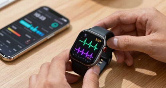
3D surgical navigation display
As an affiliate, we earn on qualifying purchases.
As an affiliate, we earn on qualifying purchases.
Conclusion
Holographic ultrasound is revolutionizing surgical practices, offering real-time, 3D imaging that enhances precision and confidence during procedures. You might worry about the technology being too complex or costly, but the benefits far outweigh these concerns. As it becomes more integrated into healthcare, you’ll see improved patient outcomes and streamlined surgeries. Embracing this innovation not only elevates surgical techniques but also fosters a new era of medical excellence, ensuring that both patients and surgeons thrive.

Wireless Convex Abdominal Ultrasound Probe – Handheld Portable Scanner & Medical Training Device for Clinical Detection Practice, Teaching Demos in Medical School and Skills Lab
【Wireless & Portable Design】 This handheld ultrasound scanner operates completely wirelessly, freeing you from cumbersome cables. Its portable…
As an affiliate, we earn on qualifying purchases.
As an affiliate, we earn on qualifying purchases.
holographic ultrasound for surgery
As an affiliate, we earn on qualifying purchases.
As an affiliate, we earn on qualifying purchases.